
Lavoro pubblicato sulla rivista “Diseases”. 2025 Jan 30;13(2):36.
doi: 10.3390/diseases13020036.
✅ Sfondo/obiettivi:
SARS-CoV-2 provoca stress ossidativo acuto nei polmoni tramite citochine, mediatori infiammatori e fattori apoptotici, che potrebbero causare lesioni alveolari seguite da grave sindrome respiratoria durante l’infezione da COVID-19.
La mancanza di particolari antivirali per SARS-CoV-2 ha aperto nuove strade di medicina complementare e non convenzionale come potenziale rimedio.
Lo studio attuale ha esplorato il ruolo meccanicistico della formulazione ultra-diluita di Eupatorium perfoliatum (UDE) contro lo stress ossidativo mediato dalla proteina S ricombinante SARS-CoV-2 e la mitocondropatia.
✅ Metodi:
La linea cellulare e i topi BALB/c sono stati utilizzati per segnalare che la proteina SARS-CoV-2 S ha causato una risposta infiammatoria e la successiva tempesta di citochine attraverso la via NF-κB nei polmoni insieme a danni ossidativi. L’esame morfologico è stato eseguito utilizzando la colorazione DAPI e l’istologia rispettivamente per le cellule trattate e i tessuti polmonari degli animali.
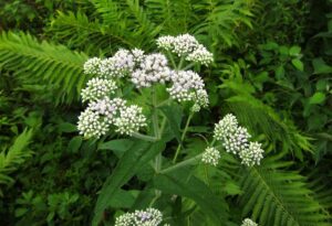
Eupatorium perfoliatum
Il meccanismo d’azione molecolare di UDE è stato studiato attraverso qRT-PCR per le espressioni genetiche di varie citochine, mediatori infiammatori e apoptotici; ELISA, immunofluorescenza, immunoistochimica e Western blot per l’espressione traslazionale delle stesse molecole testate per le espressioni genetiche; e saggi biochimici per vari enzimi e ROS.
✅ Risultati:
Il trattamento UDE (formulazione ultra-diluita di Eupatorium) ha soppresso l’infiltrazione delle cellule infiammatorie e lo stress ossidativo a livello tissutale e ha salvaguardato l’integrità mitocondriale dal danno ossidativo mediato dai radicali liberi.
Inoltre, l’UDE (formulazione ultradiluita di Eupatorium) ha svolto un ruolo diretto nel ripristino dell’omeostasi redox cellulare e nella riduzione della risposta infiammatoria sopprimendo NF-κB, IL-1β, IL-18, espressione della caspasi-1 e formazione di ROS. Inoltre, è stato proposto un meccanismo d’azione plausibile di UDE (formulazione ultra-diluita di Eupatorium) contro il danno indotto dalla proteina S.
✅ Conclusioni:
Questo studio ha descritto un nuovo approccio terapeutico contro l’iperinfiammazione mediata dalla proteina S, l’apoptosi e il danno ossidativo.
Pertanto, la formulazione ultradiluita di Eupatorium (UDE), può essere considerato come un’alternativa prospettica per combattere le conseguenze potenzialmente letali dell’infezione da SARS-CoV-2.
References:
-
Trougakos I.P., Terpos E., Alexopoulos H., Politou M., Paraskevis D., Scorilas A., Kastritis E., Andreakos E., Dimopoulos M.A. Adverse effects of COVID-19 mRNA vaccines: The spike hypothesis. Trends Mol. Med. 2022;28:542–554. doi: 10.1016/j.molmed.2022.04.007. – DOI – PMC – PubMed
-
Saini G., Aneja R. Cancer as a prospective sequela of long COVID-19. Bioessays. 2021;43:2000331. doi: 10.1002/bies.202000331. – DOI – PMC – PubMed
-
Schwartz L., Aparicio-Alonso M., Henry M., Radman M., Attal R., Bakkar A. Toxicity of the spike protein of COVID-19 is a redox shift phenomenon: A novel therapeutic approach. Free Rad. Biol. Med. 2023;206:106–110. doi: 10.1016/j.freeradbiomed.2023.05.034. – DOI – PubMed
-
Hoffmann M., Kleine-Weber H., Schroeder S., Krüger N., Herrler T., Erichsen S., Schiergens T.S., Herrler G., Wu N.H., Nitsche A., et al. SARS-CoV-2 Cell Entry Depends on ACE2 and TMPRSS2 and Is Blocked by a Clinically Proven Protease Inhibitor. Cell. 2020;181:271–280.e8. doi: 10.1016/j.cell.2020.02.052. – DOI – PMC – PubMed
-
Ramasamy S., Subbian S. Critical Determinants of Cytokine Storm and Type I Interferon Response in COVID-19 Pathogenesis. Clin. Microbiol. Rev. 2021;34:e00299-20. doi: 10.1128/CMR.00299-20. – DOI – PMC – PubMed
-
Khan S., Shafiei M.S., Longoria C., Schoggins J.W., Savani R.C., Zaki H. SARS-CoV-2 spike protein induces inflammation via TLR2-dependent activation of the NF-κB pathway. eLife. 2021;10:e68563. doi: 10.7554/eLife.68563. – DOI – PMC – PubMed
-
Huang Y., Yang C., Xu X.F., Xu W., Liu S.W. Structural and functional properties of SARS-CoV-2 spike protein: Potential antivirus drug development for COVID-19. Acta Pharmacol. Sin. 2020;41:1141–1149. doi: 10.1038/s41401-020-0485-4. – DOI – PMC – PubMed
-
Chernyak B.V., Popova E.N., Prikhodko A.S., Grebenchikov O.A., Zinovkina L.A., Zinovkin R. COVID-19 and Oxidative Stress. Biochem. Mosc. 2020;85:1543–1553. doi: 10.1134/S0006297920120068. – DOI – PMC – PubMed
-
Vollbracht C., Kraft K. Oxidative Stress and Hyper-Inflammation as Major Drivers of Severe COVID-19 and Long COVID: Implications for the Benefit of High-Dose Intravenous Vitamin C. Front. Pharmacol. 2022;13:899198. doi: 10.3389/fphar.2022.899198. – DOI – PMC – PubMed
-
Passos F.R., Heimfarth L., Monteiro B.S., Corrêa C.B., de Moura T.R., de Souza Araújo A.A., Martins-Filho P.R., Quintans-Júnior L.J., Quintans J.D. Oxidative stress and inflammatory markers in patients with COVID-19: Potential role of RAGE, HMGB1, GFAP and COX-2 in disease severity. Int. Immunopharmacol. 2022;104:108502. doi: 10.1016/j.intimp.2021.108502. – DOI – PMC – PubMed
-
Tepebaşı M.Y., İlhan İ., Temel E.N., Sancer O., Öztürk Ö. Investigation of inflammation, oxidative stress, and DNA damage in COVID-19 patients. Cell Stress. Chaperones. 2023;28:191–199. doi: 10.1007/s12192-023-01330-3. – DOI – PMC – PubMed
-
Roy A., Chakraborty R. Prophylactic and therapeutic role of catechin-loaded poly(D,L-lactic-co-glycolic acid) nanocapsules in gastric ulcer by in vitro and in vivo approach. Nanomedicine. 2023;18:197–216. doi: 10.2217/nnm-2022-0267. – DOI – PubMed
-
Ning Q., Wu D., Wang X., Xi D., Chen T., Chen G., Wang H., Lu H., Wang M., Zhu L., et al. The mechanism underlying extrapulmonary complications of the coronavirus disease 2019 and its therapeutic implication. Sig. Transduct. Target. Ther. 2022;7:57. doi: 10.1038/s41392-022-00907-1. – DOI – PMC – PubMed
-
Derksen A., Kühn J., Hafezi W., Sendker J., Ehrhardt C., Ludwig S., Hensel A. Antiviral activity of hydroalcoholic extract from Eupatorium perfoliatum L. against the attachment of influenza A virus. J. Ethnopharmacol. 2016;188:144–152. doi: 10.1016/j.jep.2016.05.016. – DOI – PubMed
-
Sinha M., Chakraborty U., Kool A., Chakravarti M., Das S., Ghosh S., Thakur L., Khuranna A., Nayak D., Basu B. In-vitro antiviral action of Eupatorium perfoliatum against dengue virus infection: Modulation of mTOR signaling and autophagy. J. Ethnopharmacol. 2022;10:114627. doi: 10.1016/j.jep.2021.114627. – DOI – PubMed
-
Sarkar A., Roy A., Maity M., Nayak D., Das S. Ultradiluted Eupatorium perfoliatum Alleviates DENV-induced Fibrosis By Regulation of TGFβ1, MMP-9 and Interferons. Future Virol. 2023;18:767–782. doi: 10.2217/fvl-2023-0121. – DOI
-
Roy A., Sarkar A., Nayak D., Das S. Ultradiluted SARS-CoV-2 Spike Protein mitigates hyperinflammation in lung via ferritin and MMP-9 regulation in BALB/c mice. Virus Res. 2023;329:199091. doi: 10.1016/j.virusres.2023.199091. – DOI – PMC – PubMed
-
Roy A., Sarkar A., Ghosh P.M., Parveen S., Nayak D., Das S. Safety Evaluation of Arsenicum album 6C in Cell Lines and BALB/c Mice Through Toxicity, Proliferation and Apoptosis Studies. Proc. Zool. Soc. 2023;76:123–133. doi: 10.1007/s12595-023-00476-3. – DOI
-
Fukushi M., Ito T., Oka T., Kitazawa T., Miyoshi-Akiyama T., Kirikae T., Yamashita M., Kudo K. Serial histopathological examination of the lungs of mice infected with influenza A virus PR8 strain. PLoS ONE. 2011;6:e21207. doi: 10.1371/journal.pone.0021207. – DOI – PMC – PubMed
-
Ramos-Martínez A.G., Enciso-Moreno J.A., Espinosa-Ayala I., Mata-Espinoza D., Rivas-Santiago B., Trujillo-Paez V., Monárrez-Espino J., Hernández-Pando R., Serrano C.J. Expression kinetics of metalloproteinases and their tissue inhibitors in experimental murine pulmonary tuberculosis. Exp. Lung Res. 2015;41:1–11. doi: 10.3109/01902148.2014.956946. – DOI – PubMed
-
Chiswick E.L., Duffy E., Japp B., Remick D. Detection and quantification of cytokines and other biomarkers. Methods Mol. Biol. 2012;844:15–30. doi: 10.1007/978-1-61779-527-5_2. – DOI – PMC – PubMed
-
Ghosh S., Dungdung S.R., Chowdhury S.T., Mandal A.K., Sarkar S., Ghosh D., Das N. Encapsulation of the flavonoid quercetin with an arsenic chelator into nanocapsules enables the simultaneous delivery of hydrophobic and hydrophilic drugs with a synergistic effect against chronic arsenic accumulation and oxidative stress. Free Radic. Biol. Med. 2011;51:1893–1902. doi: 10.1016/j.freeradbiomed.2011.08.019. – DOI – PubMed
-
Elingold I., Isollabella M.P., Casanova M.B., Celentano A.M., Pérez C., Cabrera J.L., Diez R.A., Dubin M. Mitochondrial toxicity and antioxidant activity of a prenylated flavonoid isolated from Dalea elegans. Chem. Biol. Interact. 2008;171:294–305. doi: 10.1016/j.cbi.2007.10.005. – DOI – PubMed
-
Glende E.A., Jr., Recknagel R.O. Spectrophotometric detection of lipid conjugated dienes. Methods Enzymol. 1984;105:331–337. doi: 10.1016/s0076-6879(84)05043-6. – DOI – PubMed
-
Lowry O., Rosebrough N., Farr A.L., Randall R. Protein measurement with the Folin phenol reagent. J. Biol. Chem. 1951;193:265–275. doi: 10.1016/S0021-9258(19)52451-6. – DOI – PubMed
-
Pulli B., Ali M., Forghani R., Schob S., Hsieh K.L., Wojtkiewicz G., Linnoila J.J., Chen J.W. Measuring myeloperoxidase activity in biological samples. PLoS ONE. 2013;58:e67976. doi: 10.1371/journal.pone.0067976. – DOI – PMC – PubMed
-
Klopfleisch R. Multiparametric and semiquantitative scoring systems for the evaluation of mouse model histopathology—A systematic review. BMC Vet. Res. 2013;9:123. doi: 10.1186/1746-6148-9-123. – DOI – PMC – PubMed
-
Meyerholz D.K., Beck A.P. Histopathologic Evaluation and Scoring of Viral Lung Infection. Methods Mol. Biol. 2020;2099:205–220. doi: 10.1007/978-1-0716-0211-9_16. – DOI – PMC – PubMed
-
Leist S.R., Schäfer A., Martinez D.R. Cell and animal models of SARS-CoV-2 pathogenesis and immunity. Dis. Model. Mech. 2020;13:dmm046581. doi: 10.1242/dmm.046581. – DOI – PMC – PubMed
-
Wang Z., Li S., Huang B. Alveolar macrophages: Achilles’ heel of SARS-CoV-2 infection. Sig Transduct. Target. Ther. 2022;7:242. doi: 10.1038/s41392-022-01106-8. – DOI – PMC – PubMed
-
Costela-Ruiz V.J., Illescas-Monte R., Puerta-Puerta J.M., Ruiz C., Melguizo-Rodríguez L. SARS-CoV-2 infection: The role of cytokines in COVID-19 disease. Cytokine Growth Factor. Rev. 2020;54:62–75. doi: 10.1016/j.cytogfr.2020.06.001. – DOI – PMC – PubMed
-
Kabe Y., Ando K., Hirao S., Yoshida M., Handa H. Redox regulation of NF-kappaB activation: Distinct redox regulation between the cytoplasm and the nucleus. Antioxid. Redox Signal. 2005;7:395–403. doi: 10.1089/ars.2005.7.395. – DOI – PubMed
-
Liu C., Chu D., Kalantar-Zadeh K., George J., Young H.A., Liu G. Cytokines: From Clinical Significance to Quantification. Adv. Sci. 2021;8:e2004433. doi: 10.1002/advs.202004433. – DOI – PMC – PubMed
-
Zheng M., Karki R., Williams E.P., Yang D., Fitzpatrick E., Vogel P., Jonsson C., Kanneganti T. TLR2 senses the SARS-CoV-2 envelope protein to produce inflammatory cytokines. Nat. Immunol. 2021;22:829–838. doi: 10.1038/s41590-021-00937-x. – DOI – PMC – PubMed
-
Cao X., Tian Y., Nguyen V., Zhang Y., Gao C., Yin R., Carver W., Fan D., Albrecht H., Cui T., et al. Spike Protein of SARS-CoV-2 Activates Macrophages and Contributes to Induction of Acute Lung Inflammations in Mice. FASEB J. 2021;35:e21801. doi: 10.1096/fj.202002742RR. – DOI – PMC – PubMed
-
Hardy E., Fernandez-Patron C. Targeting MMP-Regulation of Inflammation to Increase Metabolic Tolerance to COVID-19 Pathologies: A Hypothesis. Biomolecules. 2021;11:390. doi: 10.3390/biom11030390. – DOI – PMC – PubMed
-
Koppula S., Kumar H., Kim I.S., Choi D.K. Reactive oxygen species and inhibitors of inflammatory enzymes, NADPH oxidase, and iNOS in experimental models of Parkinson’s disease. Mediators Inflamm. 2012;2012:823902. doi: 10.1155/2012/823902. – DOI – PMC – PubMed
-
Meissner F., Molawi K., Zychlinsky A. Superoxide dismutase 1 regulates caspase-1 and endotoxic shock. Nat. Immunol. 2008;9:866–872. doi: 10.1038/ni.1633. – DOI – PubMed
-
Van Opdenbosch N., Lamkanfi M. Caspases in Cell Death, Inflammation, and Disease. Immunity. 2019;50:1352–1364. doi: 10.1016/j.immuni.2019.05.020. – DOI – PMC – PubMed
-
Yu J., Nagasu H., Murakami T., Hoang H., Broderick L., Hoffman H.M., Horng T. Inflammasome activation leads to Caspase-1-dependent mitochondrial damage and block of mitophagy. Proc. Natl. Acad. Sci. USA. 2014;111:15514–15519. doi: 10.1073/pnas.1414859111. – DOI – PMC – PubMed
-
Kellner M., Noonepalle S., Lu Q., Srivastava A., Zemskov E., Black S.M. ROS Signaling in the Pathogenesis of Acute Lung Injury (ALI) and Acute Respiratory Distress Syndrome (ARDS) Adv. Exp. Med. Biol. 2017;967:105–137. doi: 10.1007/978-3-319-63245-2_8. – DOI – PMC – PubMed
-
Kale J., Osterlund E.J., Andrews D.W. BCL-2 family proteins: Changing partners in the dance towards death. Cell Death Differ. 2018;25:65–80. doi: 10.1038/cdd.2017.186. – DOI – PMC – PubMed
-
Kesavardhana S., Malireddi R.S., Kanneganti T.D. Caspases in Cell Death, Inflammation, and Pyroptosis. Annul. Rev. Immunol. 2020;38:567–595. doi: 10.1146/annurev-immunol-073119-095439. – DOI – PMC – PubMed
Comunicazione a cura di:
Centro di Salute Globale SIMOH
Scuola Italiana di Medicina Omeopatica Hahnemanniana
Via Giovanni Miani, 8 – 00154 Roma
www.omeopatiasimoh.org – info@omeopatiasimoh.net